


Liver Segmentation using Unet Medical Image Analysis in Python Projects
Liver Segmentation using Unet Medical Image Analysis in Python Projects

Liver Segmentation using Unet Medical Image Analysis in Python Projects
Abstract
Liver segmentation from medical images is a critical step in diagnosing liver diseases, planning surgical interventions, and monitoring treatment outcomes. The project Liver Segmentation using U-Net for Medical Image Analysis in Python Projects focuses on developing an automated system that accurately segments liver regions from CT or MRI scans using deep learning. Python is used as the development platform because of its robust libraries for medical image processing, deep learning, and visualization, including TensorFlow, Keras, OpenCV, SimpleITK, and NumPy. The system preprocesses medical images to remove noise, normalize intensities, and enhance contrast, then applies a U-Net convolutional neural network architecture for precise pixel-level segmentation. Automated liver segmentation improves diagnostic efficiency, reduces manual effort, and supports clinicians in treatment planning and liver disease management.
Existing System
Existing methods for liver segmentation often rely on manual annotation by radiologists or semi-automated image processing techniques. Manual segmentation is time-consuming, subjective, and prone to inter-observer variability, while traditional methods such as thresholding, region growing, or edge detection may fail with complex liver shapes, low contrast, or overlapping organs. Some conventional machine learning approaches using handcrafted features have been attempted, but they lack the ability to generalize across diverse datasets and cannot capture complex anatomical structures effectively. Consequently, existing systems are limited in accuracy, robustness, and scalability for clinical applications.
Proposed System
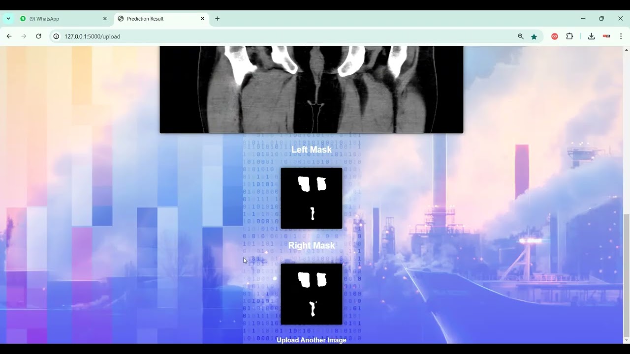
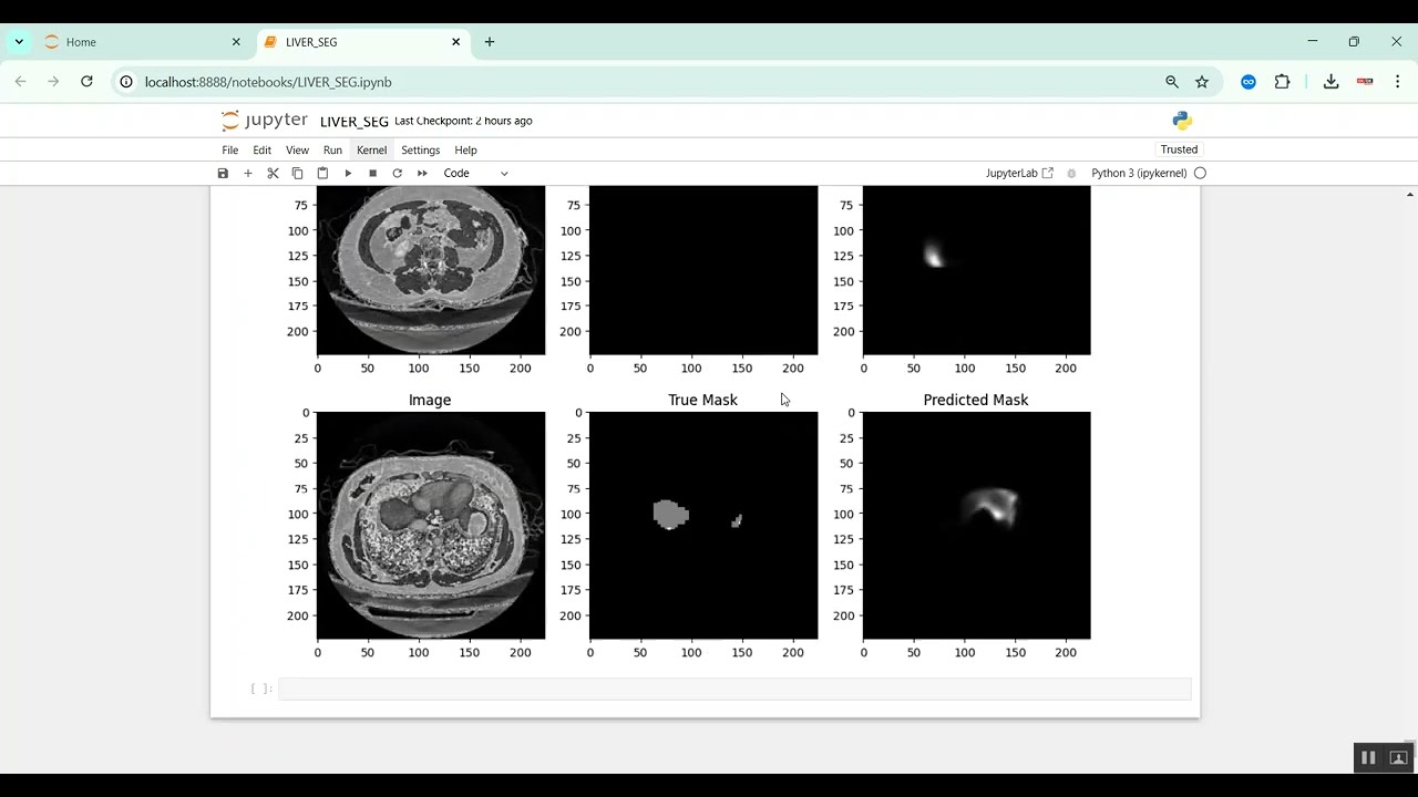
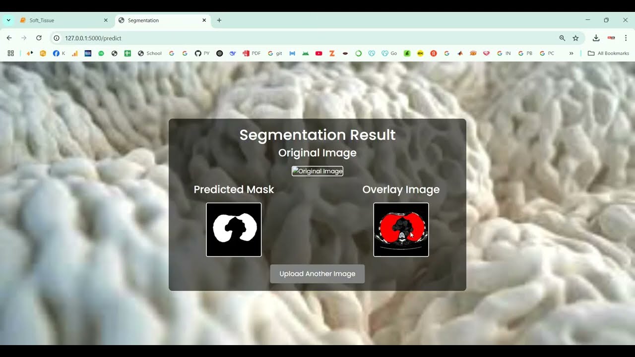
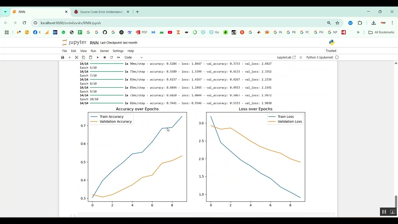
The proposed system introduces a Python-based deep learning framework for automated liver segmentation using U-Net architecture. Preprocessing steps include noise reduction, intensity normalization, resizing, and contrast enhancement to improve image quality. The U-Net model is trained on annotated CT/MRI datasets to perform pixel-level segmentation, accurately distinguishing liver tissues from surrounding organs and structures. Data augmentation techniques, such as rotation, flipping, scaling, and elastic transformations, are applied to improve model robustness and generalization. The system outputs segmented liver masks overlayed on the original images for visualization and further clinical analysis. Performance evaluation is conducted using metrics such as Dice coefficient, Intersection over Union (IoU), precision, and recall. This approach provides accurate, automated, and reliable liver segmentation, facilitating early diagnosis, treatment planning, and monitoring of liver diseases.
What's Your Reaction
 Like
0
Like
0
 Dislike
0
Dislike
0
 Love
0
Love
0
 Funny
0
Funny
0
 Angry
0
Angry
0
 Sad
0
Sad
0
 Wow
0
Wow
0